Embryo Grading and Assessment
WHAT IS EMBRYO GRADING
Embryo grading helps assess the quality and stage of embryos during IVF. Embryologists check the number of cells and how uniform their size and shape are. Higher-quality embryos may have a better chance of leading to pregnancy, but grading of embryo is just one factor in the process
STANDARD EMBRYO DEVELOPMENT TIMELINE
Day 0: Egg maturity is assessed and mature eggs are fertilized
Day 1: Fertilization assessment
Day 2: Embryo is between 2-4 cells
Day 3: Embryo is between 6-8 cells
Day 4: Embryo is at Compacting (8-16 cells) or Morula (16-32 cells) stages
Day 5-7: Embryo is at Cavitation stage or becomes a Blastocyst (100-200 cells)
Day 1 - Zygote Fertilization Grading
On Day 1 of development, the fertilized zygote is assessed to confirm successful fertilization. The zygote stage begins at fertilization when the sperm and egg fuse to form a single-cell organism. This stage lasts for about 24 hours, during which the zygote undergoes its first cell division. This stage is crucial for the grading of embryos, which helps determine their quality and potential for successful outcomes. Embryo grading considers the presence of pronuclei to evaluate viability.
Day 1 Stages
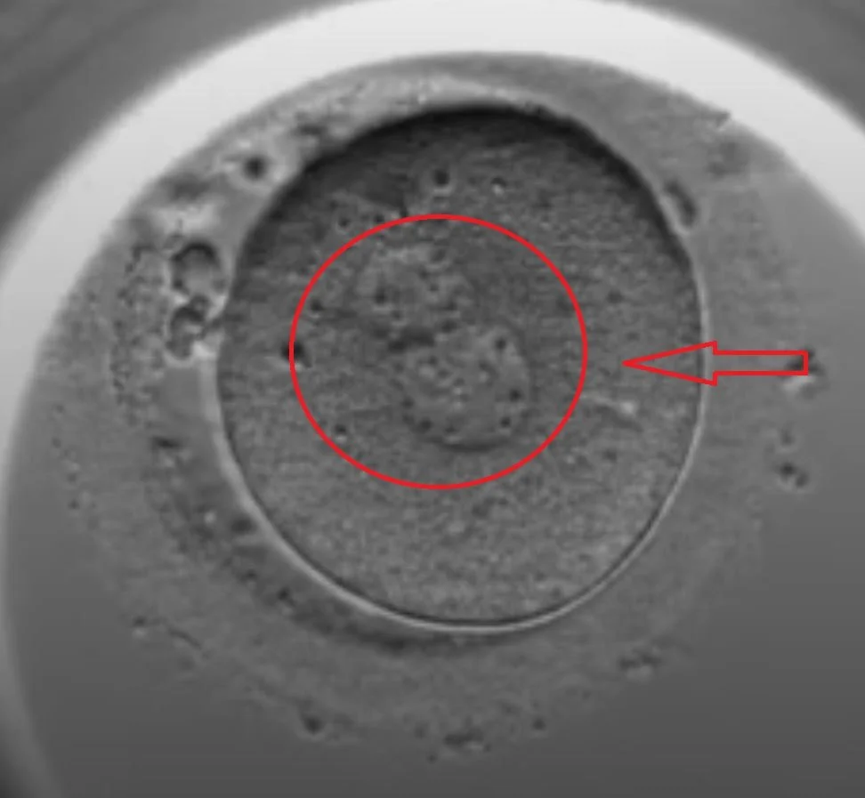
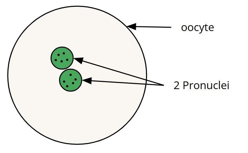
2PN (Two Pronuclei): Successful fertilization. The egg and sperm pronuclei have merged, and the zygote is now ready for the first cell division
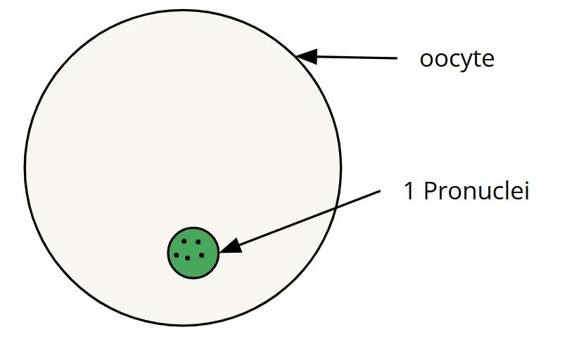
1PN (One Pronuclei): This indicates a potential fertilization issue, as only one pronucleus is visible, suggesting a failure of sperm and egg fusion
0PN (No Pronucleus): This indicates that fertilization did not occur, and the egg is unlikely to develop further. Some labs may call this PB stage because only a polar body is visible
Multinucleated Zygote: Rare, but this can indicate abnormal fertilization, where the zygote has more than two pronuclei, typically leading to developmental failure. Some labs may call this 3PN, 4PN, or Multi-PN stage
Day 1 Fertilization Stages and Assessment
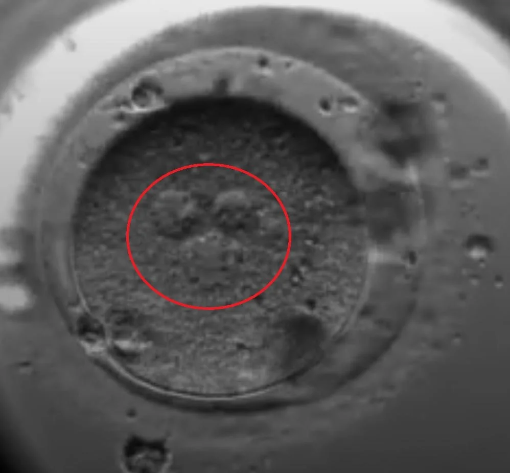
Two Pronuclei (2PN)
Successful Fertilization
The egg and sperm pronuclei have merged, and the zygote is now ready for the first cell division
One Pronuclei (1PN)
Abnormal Fertilization
Fertilization has occurred, but only one pronucleus is visible instead of the expected two

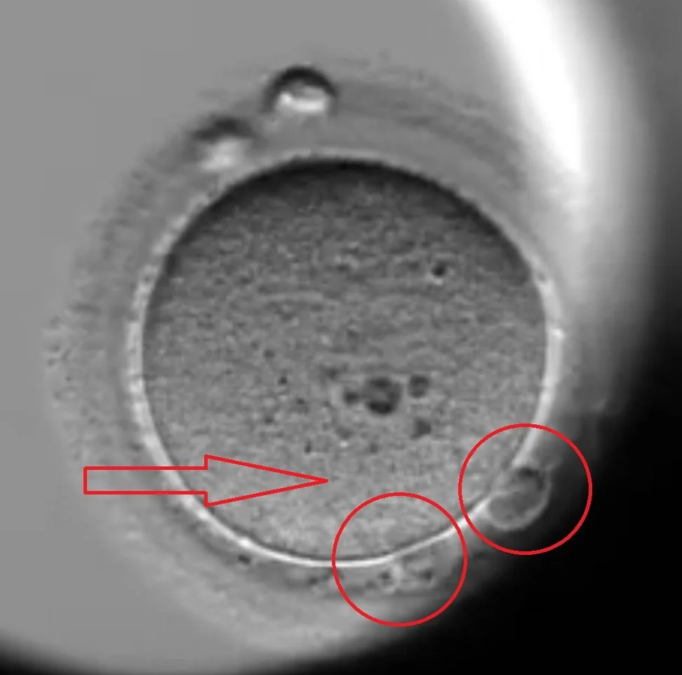
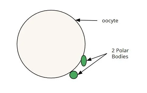
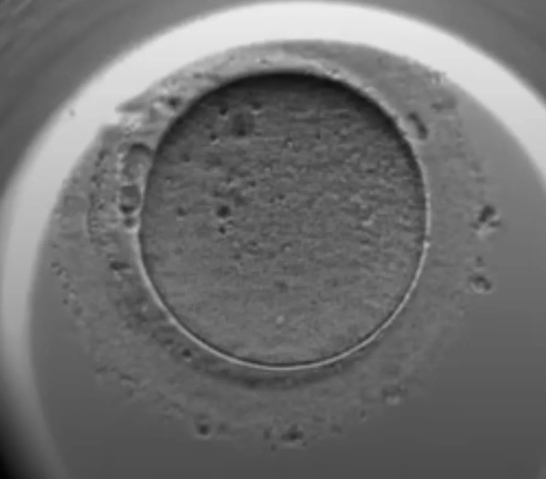
2 Polar Bodies (2PB)
Possible Fertilization Failure or Abnormal Activation
The presence of two polar bodies without visible pronuclei suggests that the oocyte may have extruded the second polar body but failed to form detectable pronuclei.
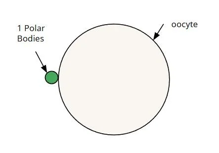
0PN or PB
Failed Fertilization.
The presence of a single polar body without pronuclei suggests the oocyte failed to fertilize. This could be from failed sperm entry or issues with oocyte maturation
3PN or Multi-PN
Abnormal Fertilization.
The presence of three or more pronuclei indicates abnormal fertilization, often due to polyspermy (fertilization by multiple sperm) or failed extrusion of the second polar body

Day 2, 3, 4 - Embryo Grading
Embryo grading on Days 2, 3, and 4 helps embryologists assess the quality and development of embryos as they progress toward the blastocyst stage. The embryo grading system evaluates factors like cell number, symmetry, and fragmentation to predict implantation potential. Grading of embryos on Day 2 and Day 3 typically focuses on the number of cells and overall appearance. Towards the end of Day 3, embryologists also check for compaction formation while Day 4 assessment looks for morula formation. Embryo grading provides valuable insights and it is just one of many factors influencing IVF success. Most labs follow some version of the Gardner Embryo Grading System.
Embryo Grading Assessment
A (Top Quality):
Evenly sized blastomeres (cells)
Little to no fragmentation (0–10%)
Smooth cytoplasm with no visible abnormalities
B (Good to Fair Quality):
Slightly uneven blastomeres but still organized
Moderate fragmentation (10–25%)
Minor cytoplasmic irregularities
C (Poor Quality):
Noticeably uneven blastomeres (size differences)
High fragmentation (>25%)
Cytoplasmic abnormalities (dark, grainy, or vacuolated appearance)
Day 2, 3, 4 Stages
Day 2: Embryos typically reach 4 to 6 cells through cleavage divisions
Example - 4A means 4-cell cleavage stage embryo with perfect symmetry and no fragmentation
Day 3: Embryos usually reach 6 to 10 cells, ideally the 8-cell stage.
Towards the end of Day 3, some embryos begin compaction formation (8-16 cells), where cell boundaries start merging
Day 4: Embryos are in the compaction stage and start to enter the morula stage (16-32 cells)
Towards the end of Day 4, some embryos begin cavitation formation (50-100 cells), where a fluid-filled cavity begins forming inside the morula.
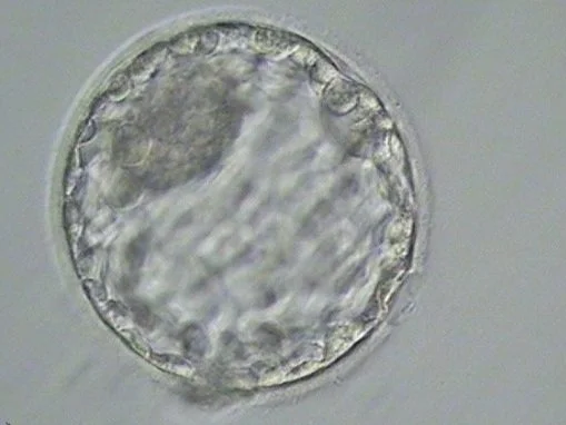
Day 5, 6, 7 - Blastocyst Grading
Blastocyst grading on Days 5, 6, and 7 is assessed using the Gardner grading scale, which evaluates expansion, inner cell mass (ICM), and trophectoderm (TE) quality. Expansion is graded from 1-6, with fully expanded and hatching blastocysts (grades 4-6) being ideal for transfer or freezing. The ICM, which develops into the fetus, is graded as A (dense, tightly packed), B (loose, less defined), or C (few, scattered cells). The TE, which forms the placenta, is similarly graded from A to C based on the number and organization of cells. High-quality blastocysts with strong ICM and TE scores are preferred for implantation potential and successful pregnancy outcomes.
Blastocyst Grading Assessment
First Character - Expansion 1-6
1 - Early Blastocyst
2 - Blastocyst
3 - Full Blastocyst
4 - Expanded Blastocyst
5 - Hatching Blastocyst
6 - Hatched Blastocyst
Second Character - Inner Cell mass (ICM) A-C
A - Good (Tightly packed ICM with many cells)
B - Fair (Loosely grouped ICM with several cells)
C - Poor (Very few cells and disorganized)
Third Character - Trophectoderm (TE) Quality A-C
A - Good (TE with many cells forming a cohesive epithelium)
B - Fair (Few cells forming a loose epithelium)
C - Poor (Very few large cells)
Blastocyst Grading Examples
4AA
6AA

5AA

3CC
